Please scroll down to watch the video.👇👇
When a large cyst squirts and oozes from a patient’s neck, it typically means the cyst has either:
-
Spontaneously ruptured, or
-
Been intentionally incised/drained by a healthcare professional.
Here’s a detailed clinical explanation of what’s happening, why it happens, and how it’s managed — including the risks involved.
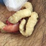
🧪 What Is Likely Happening?
A cyst is a closed sac filled with fluid, pus, keratin, or other debris. In the neck, the most common types are:
-
Epidermoid cyst (sebaceous cyst) – filled with keratin/sebum.
-
Branchial cleft cyst – congenital, near the sternocleidomastoid.
-
Infected lymph node or abscess – purulent content due to infection.
When a cyst “squirts” or “oozes,” that usually indicates increased pressure inside the cyst, forcing contents out through:
-
A spontaneous rupture, or
-
A deliberate incision by a clinician (I&D = incision and drainage).
🔍 What the Fluid Is
-
Thick, cheese-like (keratinous debris) – likely an epidermoid cyst.
-
Yellow/green, foul-smelling pus – suggests infection or abscess.
-
Clear or serosanguinous – may be a lymphatic cyst or ruptured cyst wall.
Reference: Habif TP. Clinical Dermatology. 6th Ed.
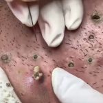
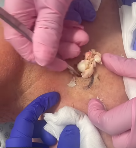
🛠️ How It’s Clinically Managed
A. Incision and Drainage (I&D)
-
Local anesthesia is used.
-
A scalpel makes a small incision over the most fluctuant point.
-
Pressure or sterile instruments are used to express contents — this may squirt if under high pressure.
-
The cavity is flushed with saline or antiseptic solution.
-
A wick or drain may be placed temporarily.
B. Culture and Antibiotics
-
If infected, pus may be sent for culture & sensitivity.
-
Empiric antibiotics started (e.g., clindamycin or amoxicillin-clavulanate) then tailored based on results.
C. Definitive Treatment (Excision)
-
After inflammation resolves, entire cyst capsule is excised to prevent recurrence.
-
If only contents are drained but wall remains, recurrence is likely.
Reference: Fitzpatrick’s Dermatology in General Medicine, 9th ed.
⚠️ Complications to Watch For
-
Recurrence (if cyst wall remains).
-
Secondary infection.
-
Scarring or sinus tract formation.
-
Abscess formation if not properly drained.
-
Cellulitis or deeper neck space infections, particularly dangerous in neck anatomy.
🧼 Home Care If Already Drained
-
Keep area clean and dry.
-
Apply antibiotic ointment (if advised).
-
Watch for redness, swelling, or fever (return to clinic if present).
-
Avoid squeezing or manipulating the area further.
📚 Clinical Reference Cases
-
Lee RA, et al. “Treatment and management of epidermoid cysts.” Am Fam Physician. 2002.
-
Nguyen T, et al. “Soft tissue infections: diagnosis and treatment.” Am Fam Physician. 2017.
A modified biopsy for squamous cell carcinoma (SCC) on the forehead involves tailoring standard biopsy techniques to accommodate the unique anatomical and cosmetic considerations of the facial region. The forehead’s prominence and the presence of underlying structures necessitate careful planning to ensure both diagnostic accuracy and optimal cosmetic outcomes.
🧠 Understanding the Need for Modification
Standard biopsy techniques—shave, punch, and excisional—are commonly employed to diagnose SCC. However, modifications are often required when dealing with facial lesions to:
-
Preserve cosmetic appearance.
-
Avoid damage to underlying structures.
-
Ensure complete and accurate sampling of the lesion.
The choice of biopsy technique depends on factors such as lesion size, depth, location, and suspicion of invasion.
🔍 Modified Biopsy Techniques for Forehead SCC
1. Modified Shave Biopsy
Indications: Raised, superficial lesions suspected to be SCC.
Technique:
-
After local anesthesia, a scalpel or razor blade is used to shave off the lesion, including a portion of the underlying dermis to ensure adequate depth.
-
Hemostasis is achieved using pressure, chemical agents, or electrocautery.
-
No sutures are typically required.
Advantages:
-
Quick and minimally invasive.
-
Preserves cosmetic appearance.
Considerations:
-
May not provide full-thickness skin samples, potentially missing deeper invasion.
Reference: American Cancer Society outlines the shave biopsy technique and its applications.
2. Modified Punch Biopsy
Indications: Lesions with uncertain depth or when full-thickness sampling is necessary.
Technique:
-
A circular blade (usually 4-6 mm) is rotated into the skin to obtain a cylindrical tissue sample that includes epidermis, dermis, and superficial subcutaneous fat.
-
The wound may be closed with a suture or allowed to heal by secondary intention.
Advantages:
-
Provides full-thickness skin samples.
-
Useful for diagnosing invasive SCC.
Considerations:
-
May require suturing, leading to a small scar.
Reference: The American Cancer Society describes the punch biopsy procedure and its indications.
3. Modified Excisional Biopsy
Indications: Small lesions where complete removal is feasible and desirable.
Technique:
-
An elliptical incision is made around the lesion, encompassing a margin of normal-appearing skin.
-
The entire lesion is removed, and the wound is closed with sutures.
Advantages:
-
Allows for complete histological assessment of the lesion and margins.
Considerations:
-
May result in a larger scar.
-
Requires careful planning to align with natural skin tension lines for optimal cosmetic results.
Reference: The American Cancer Society provides details on excisional biopsy techniques.
🧼 Post-Biopsy Care
-
Keep the biopsy site clean and dry.
-
Follow wound care instructions provided by the healthcare provider.
-
Monitor for signs of infection, such as increased redness, swelling, or discharge.
-
Attend follow-up appointments to discuss pathology results and further management.
📄 References
-
American Cancer Society. Basal and Squamous Cell Skin Cancer Tests.
-
MedlinePlus. Skin Biopsy.
-
Canadian Cancer Society. Shave Biopsy.
-